Address
Street Izvor nr. 92-96 Sector 5, Bucharest
Contact
Tel: 031.432.99.75 / 031.432.99.76
Mobil: 0755.060.992 / 0747.028.354
E-Mail: office@medicales.ro
Fax: 021.311.00.64
STB
Bus 385 – station ” Dr. Staicovici”
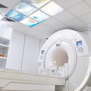
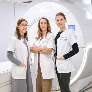
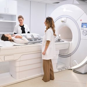
The SIGNA Voyager magnetic resonance system, produced by the General Electric company, is revolutionizing medical imaging.
Equipped with TDI (Total Digital Imaging) technology, Signa Voyager offers an exceptional quality of acquired images, clearly superior to classic magnetic resonance systems. This system increases the productivity of the medical imaging department, and its 70 cm, aperture, and antennas that use the latest technologies ensure increased comfort for patients.


SIGNA Works
The new SIGNA Works platform redefines productivity for all acquisition techniques available on the magnetic resonance system. The standard applications available on the Signa Voyager system represent an extensive set of high-quality and high-efficiency capabilities that allow obtaining the desired results for the entire examination area (neurology, body, cardio-vascular, orthopedics, senology, oncology, pediatrics).
SIGNA Voyager turns scanning into an experience without discomfort for patients. They can breathe freely thanks to the Auto Navigator technology, which is compatible with the Turbo LAVA and Turbo LAVA Flex sequences, thus allowing a complete abdominal examination, with free breathing, including in the case of sequences with contrast substance.
Using Propeller technology, the SIGNA Voyager system enables artifact-free whole-body motion acquisition, thus reducing patient stress.
Inhance Suite și 3D ASL
The Inhance Suite and 3D ASL applications allow the acquisition of angiography and perfusion images without the need to inject patients with a contrast substance, thus eliminating the risks related to this type of examination, as well as the discomfort caused to patients due to the injection.
Using the Dixon technique, available for the body, musculoskeletal, breast, and spine imaging (Ideal Flex, Lava Flex, Vibrant Flex), it is possible to acquire four different contrasts in a single scan, thus reducing the times required for examinations and, implicitly, the time spent by the patient inside the magnetic resonance system.


Computed tomography (CT) is a diagnostic imaging method that uses X-rays to create detailed images of body structures. Computed tomography allows for obtaining highly accurate images in a very short time so that a correct diagnosis can be obtained quickly.
Computerized tomography (CT) can examine any part of the body, such as the head, cervical region, chest, abdomen, pelvis, or limbs. Different organs can be investigated in detail, such as the liver, pancreas, intestines, kidneys, bladder, adrenal glands, lungs, and heart. Blood vessels, bones, and the spinal cord can also be studied.
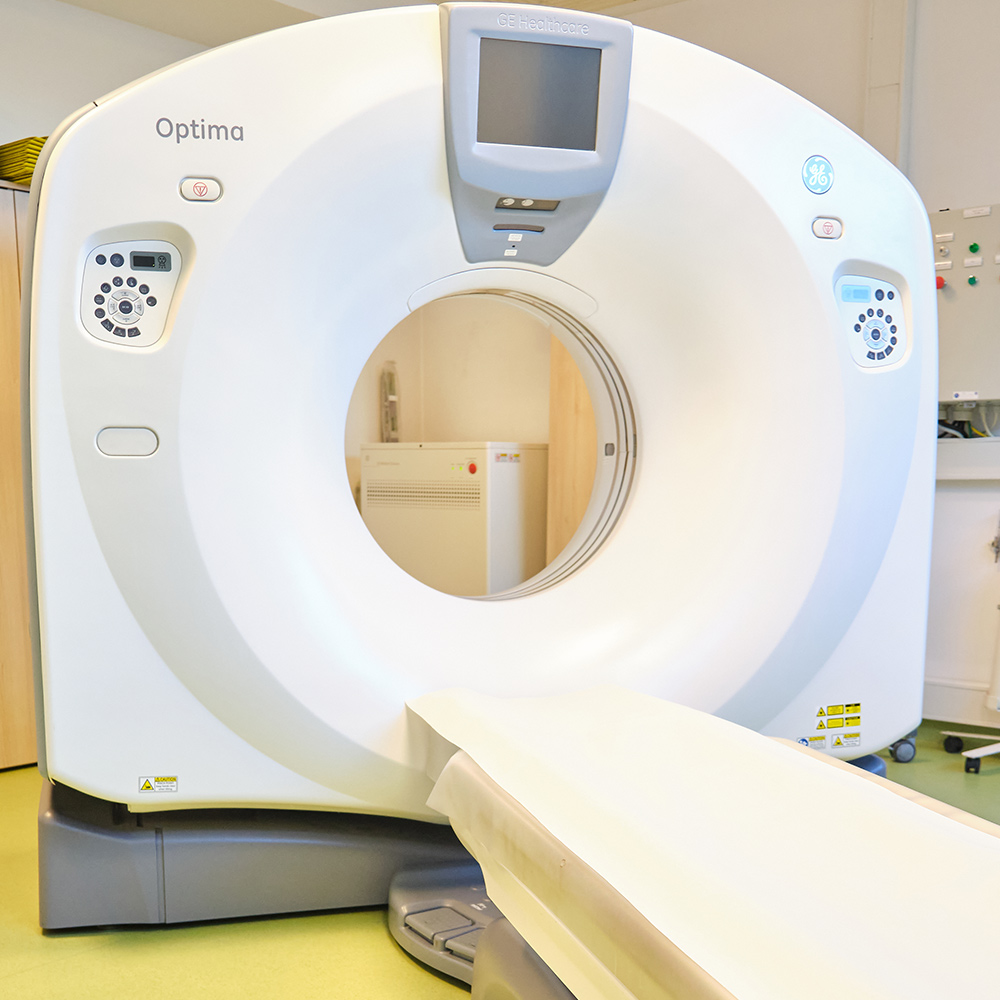
CT Optima 660
The latest generation of CT Optima 660 General Electric of 128 Slices, is the perfect solution for expanding the clinical capabilities of exploration, from simple to complex, thanks to the special scanning speed and the options with which the device is equipped. The equipment has modern technology to reduce the radiation dose administered to the patient. Options such as Smart Dose, 3D mA modulation, and Organ dose modulation help to decrease the dose administered to the patient. CT Optima 660 offers a reliable diagnosis, detecting the smallest details with a low radiation dose. The accuracy of the diagnosis is ensured by the most complex and modern post-processing station on the market, Advanced Workstation.
Bone osteodensitometry is the most modern paraclinical method by which bone mineral density (calcium content) is measured, thereby establishing the diagnosis of osteoporosis, and monitoring the evolution of the disease over time and the effectiveness of treatment.
Osteodensitometry is recommended for women after menopause, but also for men over 50 years of age, especially if they are smokers or have undergone long-term cortisone treatment.
Osteodensitometry involves scanning the body with X-rays, the radiation dose being very low, while the precision and sensitivity of the method are maximum. The investigation can be done early, from the detection of the first favorable factors, and allows the quantification of bone mineral density at the level of the entire skeleton – the “whole-body” investigation or at the level of the segments of maximum fragility (hip, spine, forearm).
Osteodensitometry has the advantage that it does not require prior preparation, and it is non-invasive and painless.


MedicalES clinics have General Electric Vivid T8 ultrasound machines – the best performing device in the field of heart ultrasounds – and General Electric Voluson E6 – the best ultrasound machine for general ultrasounds, of arteries and vessels, with dedicated software for fetal morphology.
The General Electric equipment in the MedicalES Centers allows for performing complex ultrasound explorations at the level of numerous anatomical segments:
Ultrasonographic exploration during pregnancy has reached a degree of accuracy unimaginable until a few years ago. The 3D/4D techniques that use a special type of transducer in our equipment allow on the one hand to follow the pregnancy from a young age, the early identification of certain fetal malformations, and on the other hand to reveal some morphological details on real-time that practically parents “know their fetus” before it is born:


Electromyography (EMG) is a medical procedure that consists in evaluating the function of skeletal muscles and nerves, which is extremely important in the diagnosis of peripheral nervous system disorders.

Street Izvor nr. 92-96 Sector 5, Bucharest
Tel: 031.432.99.75 / 031.432.99.76
Mobil: 0755.060.992 / 0747.028.354
E-Mail: office@medicales.ro
Fax: 021.311.00.64
Bus 385 – station ” Dr. Staicovici”
© 2024 MedicalES. All rights reserved