Burghele 1 (CT) Clinic

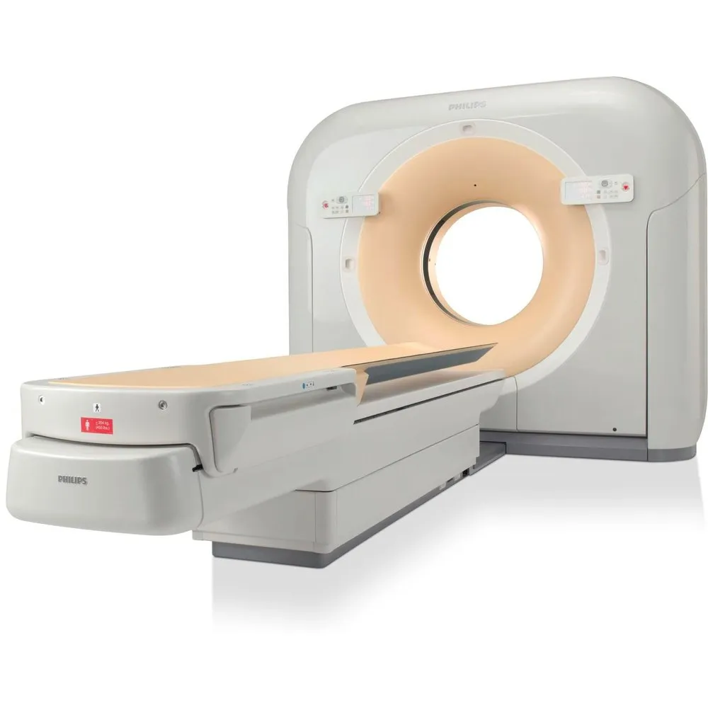
Computed Tomography MX16

Philips technology, MX16 is recognized for its speed, reliability, and clinical skills that offer patients an efficient examination and doctors a reliable diagnosis. MX 16 redefines the way of diagnosis and patient care, offering exceptional image quality, at a very low radiation dose obtained through multiple reductions and compensation techniques.

Intended for performance and patient care, the system has the lowest CT dose index, maintaining image quality at the highest standards through increased spatial resolution and unique image acquisition on a 1024 x 1024 pixel matrix.

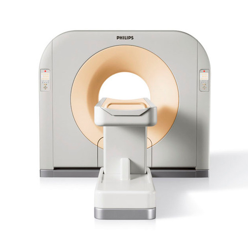
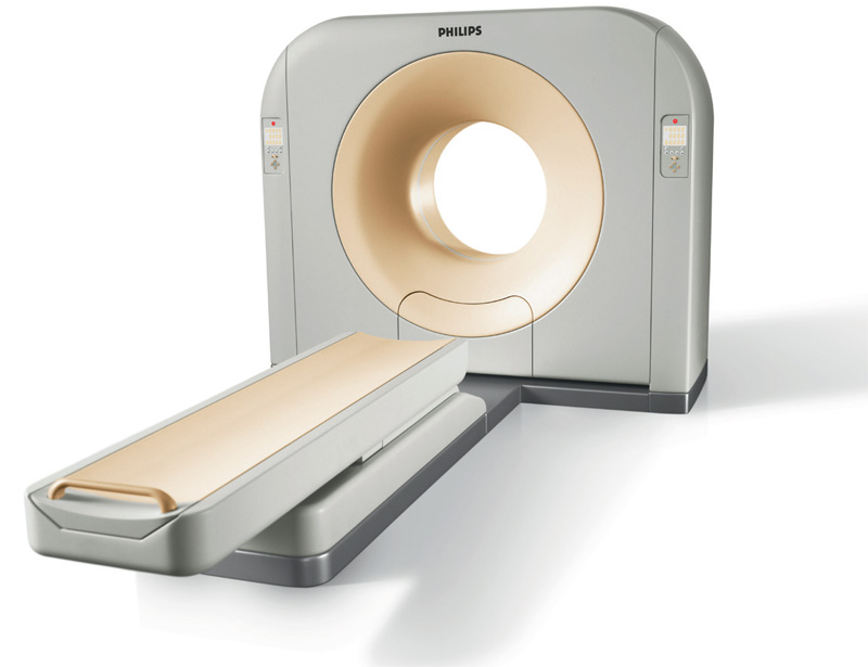
Philips MX16

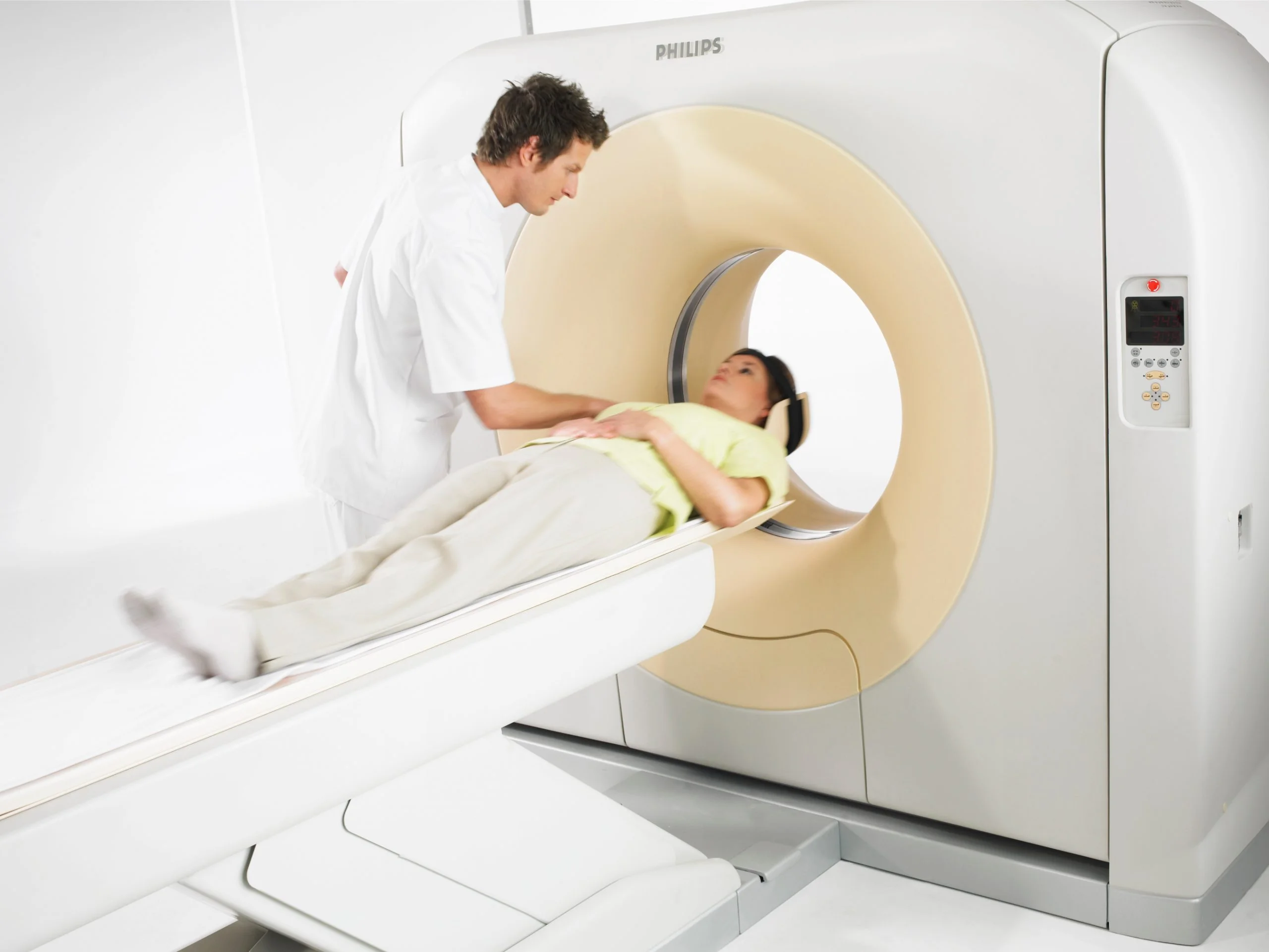
The examination table is robust and reliable, facilitating a comfortable examination, for all body types, including overweight patients. All system components, from the X-ray tube to the generator and the detector system, incorporate new Philips technologies and ambitious ideas, achieving the best results both in routine examinations and in advanced clinical applications.

Philips MX16 is a device that offers a detector coverage of 24 mm, resulting in shorter scanning and apnea times, especially favorable for pediatric examinations or anatomical structures prone to movements, such as those in the thorax-abdomen and pelvis. Exclusive technologies, such as the EvoEye algorithm and the MAR metal artifact reduction technique, offer performance and productivity.

Evo Eye

EVO EYE in computed tomography examinations, image quality is essential for diagnostic accuracy. In low-contrast images taken to visualize fine details, ring-type or band-type artifacts often appear, caused by photopenia or the polychromatic spectrum of the RX beam. The exclusive EvoEye algorithm processes and calibrates the raw data, removing artifacts and smoothing the images, without requiring additional computational time.

The results with EvoEye show an improvement in low contrast resolution from 4 mm to 2 mm at a contrast difference of 3% HU, having exceptional results in neuro studies, but also in those that require contrast substance.

MAR (Metal artifact reduction)

Artifacts caused by metal objects such as orthopedic prostheses, surgical clips, or dental fillings can be problematic in computed tomography examinations because they clutter the image with dark stripes or bands. These artifacts can thus prevent the visualization of certain anatomical structures and pathologies that could have added value to the diagnosis. The Philips MX16 system addresses this problem with the MAR metal artifact reduction algorithm. This technology isolates the effects of metal implants in the image pre-processing stage and automatically generates the conventional image and the MAR image, without requiring additional exposure.

Philips MX16

The diameter of the scanning tunnel of 70 cm ensures the increased comfort of the patient in all types of examinations, and together with the workflow efficiency, the computerized tomography system ensures the comfort and care of the patient at the highest standards, with a fast and correct diagnosis. The very short image acquisition time (complete rotation of the gantry in only 0.6 seconds) quickly provides data related to internal bleeding or other serious injuries in traumatic cases. In addition, the speed of the system reduces the apnea time to only a few seconds, increasing comfort in the case of patients with serious cardio-respiratory diseases. The exceptional quality of the images, and the reduced radiation dose, together with the efficient and fast workflow, make the MX16 computerized tomography system a high-performance equipment with high efficiency in clinical diagnosis.

Address

Sos. Panduri no. 20, Sector 5, Bucharest (in the premises of Th. Burghele Clinic)

Contact

Tel:031.436.21.82 /031.436.21.00
Mobil: 0755.060.990
E-Mail: burghele@medicales.ro

STB

122, 136, 226 – station „Spitalul Dr. Burghele”
385 – station „Soseaua Panduri”